APS/ANL
APS/ANL Heading link
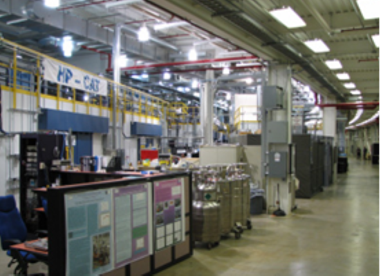
HPCAT, Advanced Photon Source
The Advanced Photon Source (APS) is a third-generation high-energy synchrotron x-ray facility at Argonne National Laboratory and is a critical resource for the proposed research program. Experiments relevant to the proposed work may be performed at several different sectors within the APS. Most of the proposed experiments will take place at the dedicated high-pressure sector, HPCAT (Sector 16), which is a principal component of our high pressure program. HPCAT is dedicated to providing state-of-the-art x-ray capabilities in extreme conditions science. The full spectrum of high-pressure x-ray experiments can be performed at this facility, including high P-T powder diffraction with laser and resistive heating, single-crystal x-ray diffraction to megabar pressures, and a variety of inelastic scattering experiments. With accurate control of the electron bunch length and spacing between bunches, the APS offers many possible operational modes ideally suited for probing dynamical processes under extreme conditions. These studies complement shock-wave studies with fourth-generation hard x-ray free-electron lasers (e.g., LCLS-II) which offer a short, brilliant pulse. APS has beamlines optimized for time-resolved, x-ray diffraction, EXAFS, along with ultrafast, high-intensity diagnostic lasers. In addition, new x-ray spectroscopy methods include nuclear resonant x-ray spectroscopy of different Mössbauer elements and resonant or non-resonant x-ray emission spectroscopy, including capabilities for studying the dramatic effects of pressure on strongly correlated d- and f- electron systems.
APS/ANL Heading link
The APS upgrade (APS-U) combined with that of HPCAT will add to the capabilities of future research on the proposed topics. On the other hand, the upgrade will require shut down of the storage ring and termination of the synchrotron x-ray experiments for a year starting in July 2022, according to current plans. Although this will preclude access to the x-ray beamline facilities that we have helped create and use during this time, we will be able to use the supporting laboratories for many experiments. During this time we will use other synchrotron x-ray sources, including the NSLS-II and PETRA III for x-ray diffraction and spectroscopy experiments.
Once the upgrade is complete, APS-U will allow experiments with improved temporal resolution by 2-3 orders of magnitude, and will enable and optimize time-resolved techniques to study kinetics and mechanisms of physical and chemical processes with a resolution ranging from sub-micro-seconds to hours. The improvement will enable further studies of the dynamics of fundamental transformations (e.g., melting, phase transitions, metastability) and transport properties (e.g., diffusivity, conductivity) under extreme conditions. HPCAT-U will also improve spatial resolution by an order of magnitude. Development of sub-µm to nm x-ray high pressure probes at HPCAT will expand the characterization of materials in an expanded P-T range, and in so doing, advance our understanding the origin of pressure-induced superconductivity, ferroelectricity, colossal magnetoresistivity, phonon softening, Fermi-surface nesting, d-electron spin pairing, f-electron delocalization, and insulator-metal and metal-insulator transitions. The development of the x-ray microscopy technique at HPCAT also creates a new tool with important implications for the proposed project.
Beamline Specific Equipment – An extensive array of instrumentation has been developed, implemented, and commissioned at HPCAT allowing a full spectrum of measurements on high-pressure samples. X-ray emission spectroscopy provides information on the filled electronic states of molecules and solids. X-ray Raman spectroscopy probes pressure-induced chemical bonding changes in light elements. Medium-resolution (0.1-1 eV) inelastic x-ray scattering spectroscopy accesses high-energy electronic phenomena, including the electronic band structure, Fermi surface, excitons, plasmons, and their dispersions. Element-specific x-ray absorption near-edge spectroscopy reveals information on oxidation state, magnetic spin, crystal-field energy, and chemical bonding changes, while extended-edge x-ray absorption fine structure reveals atomic coordination at high pressure. Resonant inelastic x-ray scattering spectroscopy probes shallow core excitations, multiplet structures, and spin-resolved electronic structure. Nuclear resonant inelastic x-ray spectroscopy provides the phonon density of states and thermodynamic information on Mössbauer elements, and nuclear resonant x-ray forward scattering is a Mössbauer absorption measurement in the time domain. These tools, coupled with x-ray diffraction, optical spectroscopy, and electromagnetic probes, hydrostatic or uniaxial pressure media, laser and resistive heating and cryogenic cooling capabilities have enabled integrated investigations of structural, vibrational, electronic, and magnetic properties at a full range of extreme conditions. A detailed description of the equipment available in each of the four stations is given below.
16-ID-D: Spectroscopy Station. The ID-D station is dedicated to x-ray spectroscopy of materials under high pressure, with a typical beam size at the sample position of 20 (v) x 50 (h) μm2. The sample stack can accommodate heavy devices such as cryostats and external heaters. An on-line spectrometer system is available for Raman and pressure measurements. The station employs a Bruker liquid nitrogen-cooled Si (111) double crystal monochromator with 2 meV energy resolution, and energy from 4.5 – 35 keV. Focusing to 4 mm (v) x 6 mm (h) μm2 is achieved with 320 mm x 400 mm IDT KB mirrors. Established techniques include:
- Nuclear Resonant Inelastic and Nuclear Forward Scattering
- Inelastic and X-Ray Raman Scattering
- Resonant and Non–Resonant X-Ray Emission Spectroscopy
- Partial Fluorescence Yield X-Ray Absorption Spectroscopy
With NRIXS at HPCAT, one can study sound velocities, magnetism and phonon density of states of materials under high pressure and at high to cryogenic temperatures. XES yields information about inner-shell and valance band electronic structure, bonding delocalization of electrons and magnetic properties in materials at high pressure. The emission spectrometer employs a spherically bent, 17-element, silicon analyzer of 1-meter radius, in near-backscattering geometry. HPCAT also has the capability for resonant emission as well as resonant inelastic x-ray scattering. X-ray Raman spectroscopy probes the nature of bonding in compounds; elements such as oxygen, nitrogen, carbon and boron have been studied at high pressures. Also, electronic excitations such as the collective plasmon excitations as well as single-particle excitations can be studied at HPCAT. This sort of inelastic x-ray scattering reveals information on the dielectric response function of materials under high pressure. At present 16-ID-D has a dedicated backscattering spectrometer with about 1 eV energy resolution. Capabilities for higher resolution spectroscopy are continuously being developed.
APS/ANL Heading link
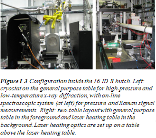
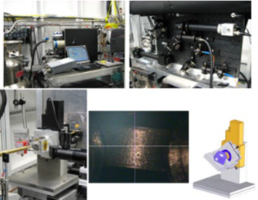
16-ID-B: Micro-Diffraction Station. The ID-B station has been configured into two major setups, one for general purpose micro-diffraction, and the other dedicated for laser heating experiments. Although the station is optimized for powder diffraction measurements in the diamond anvil cell, high pressure single crystal diffraction may also be carried out conveniently. This two-table configuration in ID-B provides the necessary space and stability for a wide variety of different micro-diffraction applications (Fig. I-3). On the laser heating table, the laser-heated DAC is combined with micro-focused synchrotron x-ray diffraction and allows the opportunity for structure-related studies of materials in-situ under ultrahigh P-T conditions. The integrated system at HPCAT combines YLF laser heating, resistive heating and micro-focused x-ray diffraction with either an imaging plate or CCD detector. The laser heating unit employs a double-sided arrangement with two identical Nd:YLF lasers (Photonics GS40, TEM01 mode, wavelength = 1053 nm), providing a total maximum output of 170 W with a power stability > 99%. For most applications, the laser is focused to a spot of ~ 30 μm; temperatures are measured on a sample area of 4×4 μm2 using an Inspectrum 300 imaging spectrograph equipped with a thermoelectric-cooled back-illuminated Hamamatsu CCD (1024 x 250 pixels), from both sides of the sample. By aligning an x-ray beam coinciding with the laser heating spot, x-ray diffraction can be carried out at simultaneous high pressures and temperatures. The typical x-ray beam size at the sample is 5×5 µm2. In addition to these state-of-the-art techniques, control systems have been developed for easy, efficient and safe operations. This system has been used for x-ray diffraction studies of a wide range of materials to over 200 GPa and above 3000 K.
The general purpose table is designed for angle dispersive diffraction measurements with a monochromatic beam, and is capable of holding bulky and heavy equipment (e.g. cryostat, graphite resistive heating assembly, large high-pressure cells) and with minimum space restrictions. The open structure and flexibility of the general purpose table allows easy modifications for performing especially demanding experiments (e.g. single crystal measurements) and the development of new techniques (e.g. high-resolution diffraction with point detectors). Double-diaphragm and piezoelectric pressure control is available for time-resolved diffraction experiments. The standard equipment on the general purpose table includes:
- A pair of 200 mm KB mirrors with a cleanup pinhole, providing a clean pseudo-Gaussian beam of 6×6 μm2 at FWHM. On demand the beam can be focused past the sample position or on the detector plane to minimize divergence or increase resolution, or just defocused for uniform sample illumination. For experiments requiring a large, low-divergence, unfocused beam the KB mirrors can be moved away.
- A heavy-duty sample stack with ample traveling range and high load capacity of 100+ kg. The sample stack has four linear and two rotational degrees of freedom. The sample stack normally is equipped with a kinematic heavy-duty cryostat or standard DAC mounting assembly, which can easily be replaced with additional stages for specific experiments (e.g. four-circle diffractometer for single crystal measurements, or combined x-y-w stage for side diffraction experiments).
- Two area detectors: MAR345 IP and MAR CCD. The standard sample-to-detector distance varies from 250 to 1100 mm, but can be changed beyond that. The horizontal tilt and position of the MAR345 IP can be easily changed to maximize the range of collected diffraction angles and optimize the quality of the spectra by minimizing the point spread function (by adjusting the horizontal tilt of the IP). The Pilatus detector is also available on request.
16-BM-D: Micro-Diffraction and X-Ray Absorption – The BM-D station is fully operational for micro x-ray diffraction and x-ray absorption spectroscopy (Fig. I-4). The station features a Si (111) double crystal monochromator in pseudo-channel cut mode and normally operates at 6-45 keV but can be configured to operate up to 60-70 keV on request. Focusing to 5 µm x 5 µm FWHM is achieved with 320 mm Pt-coated Si KB mirrors. Techniques commissioned on the beamline include:
- ADXD at simultaneous high P and high T or high P and low T
- Single crystal diffraction with the multigrain method
- Radial ADXD for stress-corrected cell parameters, texture and strength of materials at high pressure in the DAC that has been adapted with a membrane drive.
- X-ray absorption spectroscopy with ion chamber capability
Both external resistive heating and cryostat operations have been commissioned on the beamline.
APS/ANL Heading link
BM-B: Micro-Diffraction with Polychromatic Beam – The BM-B station is dedicated for the use of a white beam with third generation characteristics. A channel-cut Si (111) monochomator delivers a focused beam at 5-70 keV with a focus of 5 µm x 5 µm FWHM using a 200 mm Rh-coated Si KB mirror. Both white-beam Laue diffraction with the DAC and studies with the Paris-Edinburgh press have been commissioned. Liquid viscosity measurements with white-beam radiography and ultrasonic elastic wave velocity measurements can also be performed. The instrument allows changing the distance between the focusing optics and sample, with flexible focused beam sizes from 5 μm to 15 μm or larger depending on experimental requirements. Special features of the beamline include:
- Five-dimensional remote control for pinhole and tip (x, y, z, pitch and yaw)
- Gas driven membrane control of pressure for diamond anvil cells
- Low temperature experiments to liquid He temperature
- Point-detector (Ge detector) and area-detector (MarCCD) operation
- Automatic liquid nitrogen refill system for Ge detector
- Paris-Edinburgh type large volume press with resistive heating capacity (PE anvils, boron-epoxy gaskets, cylindrical graphite heater, 8V-220A power supply; temperature and pressure range up to 1300 K under 10 GPa or 2200 K under 7 GPa)
Two auxillary laboratories at HPCAT house a fully-equipped high-pressure facility that includes sample loading and characterization, complementing the main HPCAT laboratories at Sector 16. Apart from microscopes for aligning diamond cells, diamond polishing and shaping facilities, fume hood, gas loading system capable of loading H2 (in addition to a number of other inert gases), specialized coil winding for magnetic measurements, DC and AC conductivity measurement systems, multi-wavelength Raman spectroscopy system (660 nm, 488 nm and 532 nm) equipped with ultra narrow-band notch filters make these laboratories a self-contained facility for training and ex-situ measurements. These facilities complement those available at UIC and are available for use through the UIC-HPCAT MOU referred to above.